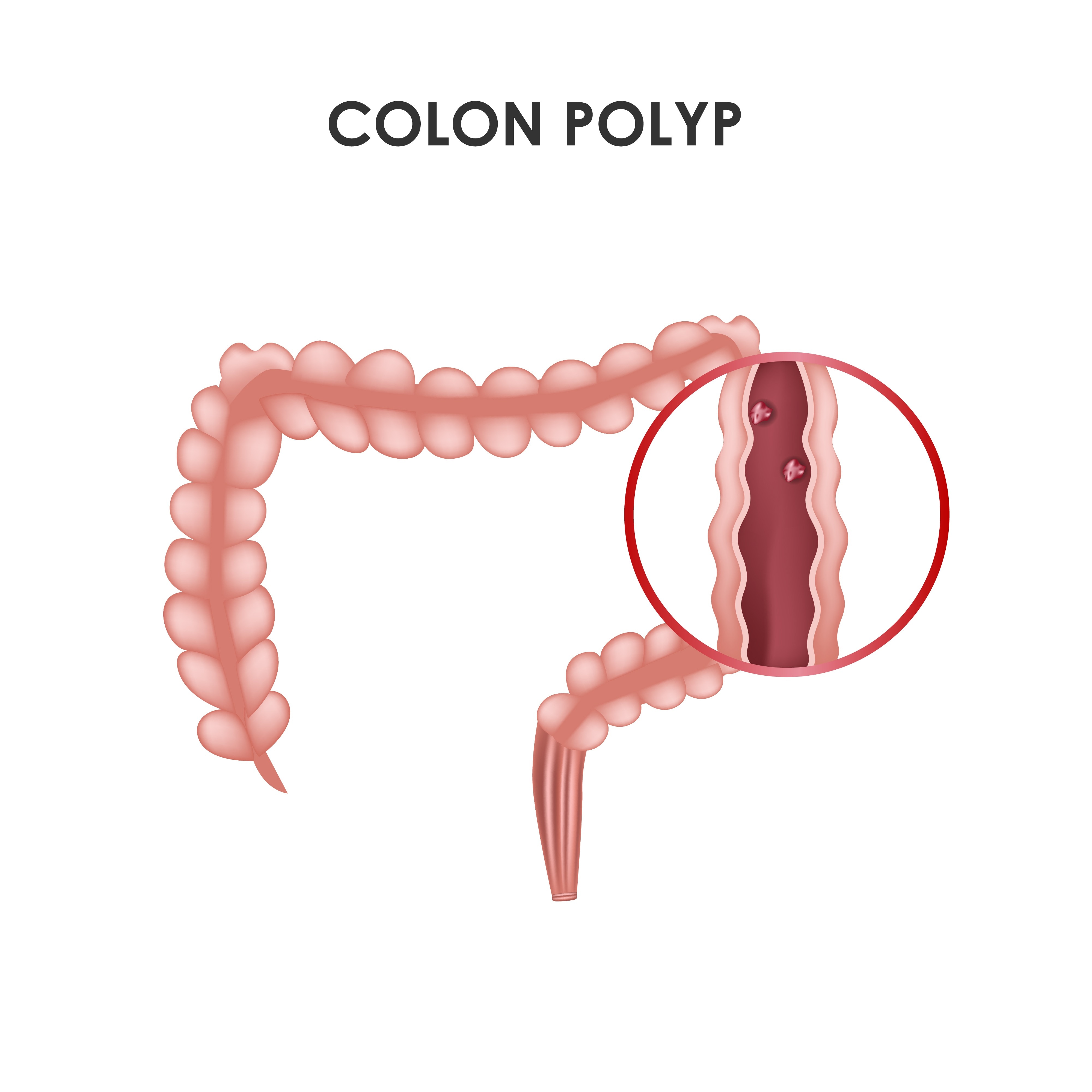
A colonic polyp can sit in the bowel without causing pain, bleeding or any noticeable change, yet some types can gradually develop into colorectal cancer. By understanding polyp types, screening choices, warning symptoms, removal methods and pathology results, you can make sense of why a finding matters and what questions to ask next.
Key takeaways
- Most colonic polyps cause no symptoms, so screening can find them before complications.
- Earlier screening matters with family history, inflammatory bowel disease or concerning symptoms.
- Colonoscopy removes polyps during the same examination; stool tests and CT scans cannot.
- Pathology results determine when your next colon examination is due.
What a Colonic Polyp Is—and Why a Silent Finding Can Matter
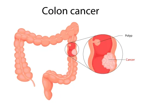
A colonic polyp is a growth from the inner lining of the large intestine. Most cause no pain, bleeding or change in bowel habits, so feeling well does not prove that a polyp is harmless.
- An adenomatous polyp, or adenoma, can develop into colorectal cancer, especially when it is larger or contains villous tissue or high-grade dysplasia.
- A serrated polyp includes a sessile serrated lesion (SSL). An SSL can be flat, pale and partly hidden behind a fold, making it harder to recognise during colonoscopy; some become cancer precursors.
- A hyperplastic polyp is usually small and located in the rectum or sigmoid colon. These distal lesions generally have little malignant potential, although not every serrated-appearing lesion is a simple hyperplastic polyp.
A doctor cannot reliably identify a polyp’s type or cancer risk by appearance alone. Removal and histopathologic examination show whether it contains dysplasia, villous architecture, invasive cancer or involved margins. That is why removing an adenomatous polyp is preventive, not merely diagnostic: it eliminates tissue that could progress over time.
Seek medical evaluation for rectal bleeding, a persistent change in bowel habits or unexplained iron-deficiency anaemia rather than waiting for routine screening. A symptom-free finding still deserves follow-up because the next step depends on the pathology report and whether removal was complete.
How Polyps Become Riskier and Who Needs Earlier Screening
Most colorectal cancers develop through the adenoma-carcinoma sequence: a conventional adenoma accumulates cellular changes, enlarges, develops advanced dysplasia, and can invade. The serrated pathway follows different molecular changes; sessile serrated lesions can be flat, subtle, and precancerous, while small distal hyperplastic polyps usually have little malignant potential.
Risk rises with polyp size, number of polyps, microscopic features, and incomplete removal. Removing adenomas during colonoscopy is preventive: the National Polyp Study associated removal with an estimated 76%–90% reduction in expected colorectal-cancer incidence during follow-up.
Screening timing depends on your risk category:
- Average-risk adults: Begin at age 45. Continue through age 75; from 76 to 85, decide with a clinician based on health, prior screening, and preferences. Options include colonoscopy, stool-based tests, and other approved screening tests.
- Family-related risk: If a parent, sibling, or child had colorectal cancer or an advanced polyp, ask about colonoscopy at age 40 or 10 years before the relative’s diagnosis, whichever comes first. A five-year interval is commonly used when family history warrants colonoscopy.
- Inherited risk: Lynch syndrome, familial adenomatous polyposis, and other syndromes require genetic and specialist planning, often beginning before age 40 and sometimes during adolescence.
- Inflammatory bowel disease: Long-standing ulcerative colitis or Crohn’s colitis generally requires surveillance colonoscopy beginning about eight years after extensive colitis starts, with intervals based on findings and inflammation.
Choosing Between Colonoscopy, Stool Tests and CT Colonography
The key difference is what each test can see. Colonoscopy examines the entire lining directly and allows the clinician to remove or sample a polyp during the same procedure. Stool tests look for signs shed into stool, while CT colonography creates images of the colon but cannot remove a lesion.
| Test | What it detects and misses | What a result leads to |
|---|---|---|
| Colonoscopy | Polyps, cancer and other lining changes; visibility depends on bowel preparation and complete insertion | Removal or biopsy during the examination |
| faecal immunochemical test (FIT) | Hidden blood; a nonbleeding polyp can produce a normal result | A positive result requires colonoscopy |
| multitarget stool DNA test | Blood plus molecular changes linked to colorectal cancer and some advanced polyps | A positive result requires colonoscopy |
| CT colonography | Three-dimensional images of the colon; small or flat lesions can be harder to identify | A suspicious finding requires colonoscopy for removal or biopsy |
A positive stool test is not a diagnosis of cancer, but it is an unfinished screening process. Arrange the follow-up colonoscopy rather than repeating the stool test, because delaying it leaves a potentially important lesion unexamined.
A normal stool test lowers concern but does not exclude polyps: bleeding can be intermittent, and some lesions do not bleed. A colonoscopy is also not an ordinary negative examination if bowel preparation hides the lining or the scope cannot reach the entire colon.
Inadequate preparation generally calls for repeat colonoscopy within one year; an incomplete examination needs medical evaluation to choose repeat colonoscopy or CT colonography.
What Happens When a Polyp Is Removed
Size, shape and suspected depth determine the removal method. A tiny, flat lesion is often treated with cold-snare polypectomy, in which a wire loop cuts it without electric heat.
A stalked lesion may need snare polypectomy with electrocautery to control the thicker blood supply; forceps biopsy is reserved for selected tiny lesions or tissue sampling, not as a substitute for complete removal when cancer risk is possible.
- Lesions under about 10 mm: cold-snare polypectomy usually removes the entire lesion in one piece.
- Stalked or larger lesions: a snare captures the head and stalk; injection or cautery can help control bleeding.
- Flat lesions around 20 mm or larger: endoscopic mucosal resection may lift and remove the lesion, sometimes through piecemeal removal.
- A lesion with features of deep invasion: do not treat it as a routine polyp; refer for advanced assessment.
The laboratory examines the tissue under a microscope to identify an adenoma, sessile serrated lesion, hyperplastic polyp or cancer. It reports villous architecture, the degree of dysplasia, invasion into deeper layers and whether the margins contain abnormal tissue. These findings show both cancer potential and whether removal was complete.
Large lesions do not automatically require an operation. An experienced advanced endoscopist can often remove a non-invasive lesion without colectomy, while suspected deep submucosal invasion calls for surgical or oncologic evaluation.
Bleeding can occur immediately or days later, and perforation is uncommon but serious; never stop an anticoagulant or antiplatelet drug without the prescribing clinician’s instructions.
Reading the Result: Surveillance, Missed Lesions and Safety Decisions
A 7–10-year interval can follow removal of one or two tubular adenomas smaller than 10 mm, but the finding alone does not set the timetable. Histopathology determines whether the tissue is adenomatous, serrated, hyperplastic, or cancerous and records villous architecture, high-grade dysplasia, invasion, and involvement of the margin.
Common U.S. surveillance examples include:
- Three years after an adenoma measuring at least 10 mm, or one with villous architecture or high-grade dysplasia.
- One year after more than 10 adenomas.
- About six months after piecemeal removal of a large lesion, so the scar can be checked for residual tissue.
An unclear margin changes the plan. A lesion that was too large for standard snaring may require endoscopic mucosal resection (EMR), and incomplete removal needs earlier review rather than routine surveillance.
Poor bowel preparation also shortens the safe interval because fluid, stool, folds, and flat or serrated lesions can hide abnormalities. A technically difficult examination or a missed segment may justify repeating colonoscopy sooner.
Bleeding can occur immediately or days after polypectomy; perforation is uncommon but serious, with greater risk after large, cauterised, difficult, or piecemeal resections. Do not stop anticoagulants or antiplatelet drugs without the prescribing clinician. Dr. Harshad Joshi can help reconcile the pathology report, procedure quality, medication risks, and safest follow-up interval.
Related service
Endoscopy Colonoscopy ₹12000 • Used for examination of the inner lining of the LARGE INTESTINES (COLON) • BOWEL PREPARATION: dietary restrictions and bowel cleansing is done to... |
Frequently asked questions
What is a colonic polyp?
A colonic polyp is a growth from the inner lining of the large intestine. Many cause no pain, bleeding or bowel-habit changes.
Who needs earlier colorectal screening?
Earlier screening matters for people with a family history of colorectal cancer or polyps, inflammatory bowel disease, or concerning symptoms.
How do colonoscopy, stool tests and CT colonography differ?
Colonoscopy allows direct examination and polyp removal. Stool tests look for signs of colorectal disease, while CT colonography uses imaging and requires follow-up colonoscopy if a lesion is found.
What happens after a polyp is removed?
The polyp is sent for laboratory examination. Its type, size, number and removal completeness help determine the surveillance interval.