Crohn’s disease can affect any digestive-tract segment from the mouth to the anus, and inflamed areas may be separated by healthy bowel. By following the tract from top to bottom, you can connect location with likely symptoms, complications and the tests doctors use without assuming that one symptom identifies one segment.
Key takeaways
- Crohn’s disease can affect any digestive-tract segment from mouth to anus.
- Skip lesions mean diseased bowel can appear between healthy sections.
- Colonoscopy examines the colon and terminal ileum; MRI and capsule endoscopy assess small bowel.
- Location guides treatment, but tests must show inflammation depth and activity.
From mouth to anus: where Crohn’s disease can occur
Crohn’s disease can occur anywhere from the mouth to the anus, although the terminal ileum, ileocolonic region and colon are common sites. The affected areas may be separated by healthy bowel, creating “skip lesions”. These Crohn’s disease locations can produce different symptoms:
| Segment | Symptoms it may produce |
|---|---|
| Mouth | Mouth ulcers, swollen lips or oral tissue, cobblestoning and deeper linear ulcers. These findings can fluctuate with bowel activity but do not prove Crohn’s disease alone. |
| Oesophagus | Oesophageal Crohn’s disease may cause painful or difficult swallowing, chest discomfort and food sticking. |
| Stomach and duodenum | Stomach and duodenum Crohn’s disease can cause upper-abdominal pain, nausea, vomiting, early fullness and, if narrowing develops, gastric outlet obstruction. |
| Jejunum and other small bowel | Cramping, diarrhoea, weight loss and nutrient deficiencies can result from inflammation, narrowing or impaired absorption. |
| Terminal ileum | Right-lower abdominal pain and diarrhoea are common. Poor absorption of vitamin B12 or bile acids can cause deficiency or watery diarrhoea, even when the colon is not the main inflamed segment. |
| Colon and rectum | Frequent diarrhoea, urgency, rectal bleeding, abdominal pain and anaemia can occur. |
| Anus and perianal skin | Fissures, painful swelling, skin tags, abscesses, fistulas and persistent drainage may appear before or alongside intestinal symptoms. |
Location does not determine severity by itself. Fever, fatigue, weight loss and pain can reflect inflammation or complications such as strictures, fistulas, infection or malabsorption.
Small-bowel, ileal and colonic Crohn’s disease behave differently
Symptoms and complications change with the segment involved: small-bowel Crohn’s disease more often causes malabsorption or narrowing, while colonic disease more often causes urgency, bleeding and frequent stools.
| Location | Common symptoms | Important complications |
|---|---|---|
| Small intestine | Cramping, diarrhoea, bloating, fatigue and weight loss | Poor absorption, anaemia, fistulas and strictures that obstruct the bowel |
| Terminal ileum | Right-lower abdominal pain, diarrhoea and weight loss | Vitamin B12 deficiency, bile-acid diarrhoea and narrowing with nausea, vomiting, distension or reduced passage of stool and gas |
| Colon | Frequent loose stools, urgency, lower-abdominal pain and visible blood or mucus | Anaemia, severe inflammation, fistulas and increased risk of bowel narrowing |
| Perianal region | Anal pain, fissures, swollen skin tags, drainage or a tender lump | Fistulas and abscesses, which can require urgent treatment |
Terminal-ileal inflammation can cause watery diarrhoea through bile-acid malabsorption, even when the colon is not the main inflamed segment. A stricture can instead dominate the picture, producing cramping and vomiting without prominent diarrhoea.
Crohn’s colitis can cause bloody diarrhoea and urgency, but symptoms do not prove the disease is limited to the colon. Perianal Crohn’s disease can precede bowel symptoms, so persistent drainage, recurrent abscesses or painful swelling deserves assessment rather than treatment as an isolated local problem.
Fatigue, fever, weight loss and anaemia can occur at any location because of inflammation, blood loss, malabsorption, infection or medication effects. Doctors therefore match symptoms with blood tests, biopsies and small-bowel imaging.
Why location alone does not show how active or deep the disease is
A short abnormal segment does not necessarily mean mild or shallow disease: Crohn’s can be patchy, buried in the bowel wall, or already causing a mechanical complication. Skip lesions leave normal-looking bowel between inflamed areas, so a limited view can understate the disease pattern.
The visible length also says little about how far inflammation extends through the wall.
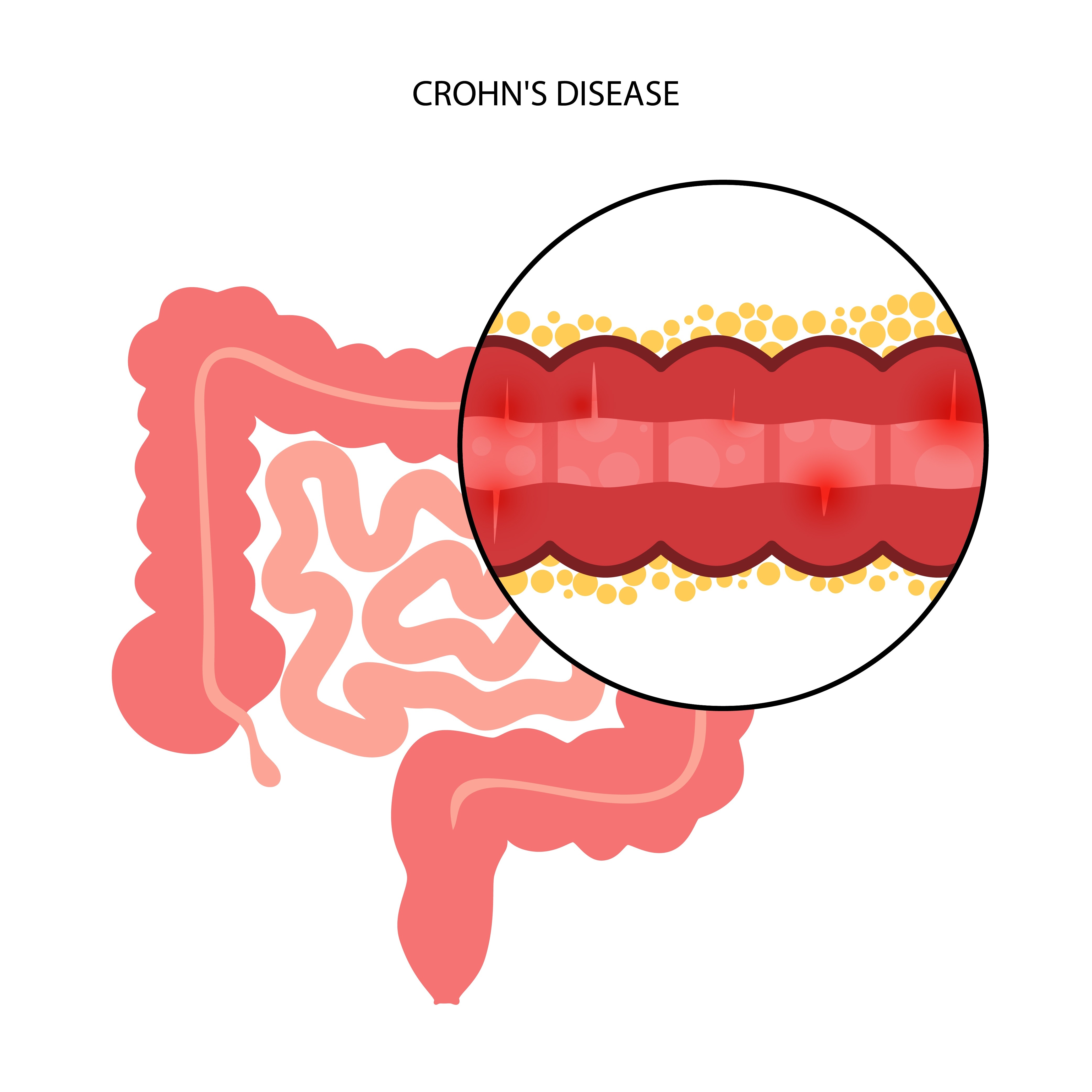
Transmural inflammation can involve the full thickness of the bowel wall, even when the surface appears only modestly abnormal. That depth can cause:
- An ileal stricture, where swelling or scarring narrows the passage.
- Intestinal obstruction, with cramping, abdominal distension, nausea, vomiting, or reduced stool and gas, even without prominent diarrhoea.
- An abscess or fistula, when inflammation extends beyond the bowel.
Symptoms do not reliably measure activity or depth. Pain, diarrhoea, fatigue, weight loss and fever can arise from different mechanisms, including inflammation, narrowing or penetrating disease. Endoscopy with biopsies examines the lining, but it cannot fully assess transmural or extra-luminal disease.
Magnetic-resonance enterography or computed-tomography enterography can show bowel segments beyond the colonoscope and identify wall thickening, strictures, abscesses and fistulas. Doctors therefore judge an apparently limited area using symptoms, objective tests and the structure of the bowel wall—not location alone.
Which investigation examines each digestive-tract segment?
Gastroscopy is the direct test for the oesophagus, stomach and duodenum. It shows ulcers, narrowing and surface inflammation, but it cannot assess most of the jejunum or ileum and does not show disease outside the bowel wall.
| Digestive-tract target | Best investigation | What it can miss |
|---|---|---|
| Upper tract: oesophagus, stomach and duodenum | Gastroscopy with biopsies | Jejunum, ileum, deeper bowel-wall disease and fistulas beyond its reach |
| Colon and terminal ileum | Colonoscopy with terminal-ileum intubation and biopsies | Narrowed segments the scope cannot pass, deeper or extra-luminal disease, and inflammation hidden between patchy lesions |
| Deeper small bowel: jejunum and ileum | Magnetic-resonance enterography; computed-tomography enterography when urgent speed matters | Mucosal detail can be less precise than direct endoscopy; CT uses ionising radiation |
| Bowel-wall complications: thickening, strictures, fistulas and abscesses | Intestinal ultrasound or cross-sectional imaging | Intestinal ultrasound can be limited by bowel gas, obesity and operator experience; small fistulas or abscesses may need MRI or CT |
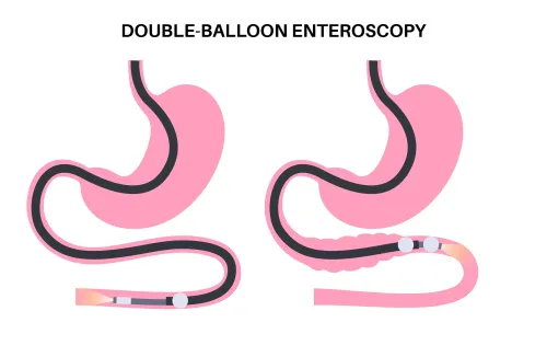
| Small-bowel surface inflammation | Capsule endoscopy | Capsule endoscopy limitations include no biopsy, no treatment and failure to pass a stricture; it can also miss disease hidden beneath normal-looking mucosa |
A normal colonoscopy does not exclude Crohn’s disease in the small bowel, and a normal surface view does not exclude transmural inflammation. Dr. Harshad Joshi can help match the test to the suspected segment, obstruction risk and question that needs answering rather than ordering every investigation at once.
Related services
Endoscopy Colonoscopy ₹12000 • Used for examination of the inner lining of the LARGE INTESTINES (COLON) • BOWEL PREPARATION: dietary restrictions and bowel cleansing is done to... |
Ultrasound ₹2500 It is a specific type of abdominal ultrasound that focuses on imaging and assessing the small intestine. View service → |
How doctors connect symptoms, urgency and treatment to the affected location
A painful, swollen area beside the anus, pus, or new drainage needs same-day assessment. It may indicate an abscess or perianal fistula requiring examination and drainage, not just cream.
Seek urgent care for any of these Crohn’s disease red flags:
- Severe or rapidly worsening abdominal pain
- Persistent vomiting or inability to keep fluids down
- A rigid or markedly swollen abdomen
- Inability to pass stool or gas
- Heavy rectal bleeding, fainting, or high fever
These signs can reflect obstruction, infection, significant bleeding, or deep inflammation. Less dramatic symptoms also need assessment when diarrhoea, pain, fatigue, weight loss, or fever persists, because location alone cannot show disease activity.
Doctors combine the symptom pattern with blood tests, stool tests, endoscopy and biopsies; small-bowel imaging adds information when a stricture, abscess, or fistula is suspected.
Treatment depends on what the inflammation is doing. Ileal disease can cause vitamin B12 deficiency or bile-acid diarrhoea, while extensive small-bowel disease can cause weight loss and malabsorption; dietetic review and targeted replacement prevent avoidable deficiencies. Colonic bleeding can produce iron-deficiency anaemia.
A stricture may require escalation of medical treatment or Crohn’s disease surgery, while an abscess needs drainage before immune-suppressing treatment. Perianal disease often needs coordinated medical and surgical care. Extraintestinal manifestations can also alter drug choice and prompt assessment outside the digestive tract.
Frequently asked questions
Where in the digestive tract can Crohn’s disease occur?
Crohn’s disease can occur from the mouth to the anus, including the small intestine, terminal ileum, ileocolonic region and colon. Diseased areas can be separated by healthy bowel, creating skip lesions.
How do small-bowel, ileal and colonic Crohn’s disease differ?
Small-bowel Crohn’s disease can cause abdominal pain, diarrhoea and nutrient deficiencies. Ileal disease can contribute to pain in the lower right abdomen, narrowing and vitamin B12 or bile-salt problems. Colonic disease more often causes diarrhoea, urgency, blood or mucus, and rectal symptoms.
Does Crohn’s disease location show how active or deep it is?
No. Location identifies where disease is found, not whether inflammation is active or how deeply it extends through the bowel wall. Endoscopy, biopsy and cross-sectional imaging help assess those features.
Which investigations examine different digestive-tract segments?
Upper endoscopy examines the oesophagus, stomach and duodenum. Colonoscopy examines the colon and terminal ileum. MR enterography or CT enterography assesses the small bowel and complications, while capsule endoscopy can view areas that standard scopes cannot reach.
How does disease location affect Crohn’s disease treatment urgency?
Doctors combine location with symptoms, inflammatory activity, complications and nutritional effects. Obstruction, abscess, fistula, significant bleeding, dehydration or severe pain requires prompt medical assessment, regardless of the affected segment.
Related services
Endoscopy Enteroscopy ₹70000 Enteroscopy is a medical procedure used to visualize and examine the small intestine, which is located between the stomach and the large intestine... |
Endoscopy ₹25000 Patient Information: Endoscopic Ultrasound (EUS) What is Endoscopic Ultrasound (EUS)? Endoscopic Ultrasound (EUS) is a minimally invasive procedure... View service → |
Endoscopy Gastroscopy ₹9000 Examination of ESOPHAGUS, STOMACH, DUODENUM • Painless: Done under mild sedation • SAFE: in experienced hands. USES: 1. |
Endoscopy ₹50000 Capsule endoscopy is a non-invasive medical procedure that involves swallowing a small, pill-sized capsule equipped with a camera and light source. View service → |